Octopus Visual Field

In 1945, Professor Hans Goldmann of the University of Berne, together with Haag-Streit, developed the Manual Goldmann perimeter. This instrument is still the reference for kinetic perimetry today and all of its capabilities and specifications are built into the Octopus 900. Furthermore, Haag-Streit has pioneered many significant innovations, including automated static perimetry, the G-Program and the direct projection system, as well as fast strategies and outstanding software for visual field analysis. Addressing all major perimetry needs, the Octopus 900 is the perimeter of choice for anyone who wants diagnostic flexibility to analyse, assess and track patients’ visual fields. It makes it possible to run standard central and peripheral visual fields with minimum test duration, seamlessly integrated into your practice environment. This supports high patient throughput and effective practice management. With its built-in reliability features, the Octopus 900 is easy-to-use and delivers results you can trust. This device is the visual field of choice used by the world renowned Wills Eye Institute.
OCT

CIRRUS HD-OCT 5000 is configured with the highest resolution visualization capabilities and sophisticated clinical applications such as Advanced RPE analysis to track retinal pigment epithelial integrity and Ganglion Cell Analysis to assess glaucomatous loss in the macula that may not be evident in the peripapillary region. The new FastTrac™ retinal tracking reduces eye motion artifacts without sacrificing patient throughput with a proprietary scan acquisition strategy, high speed 20 Hz LSO camera, and single-pass alignment scanning. This feature enables the highest resolution B-Scans to be captured in identical locations from visit to visit providing precise assessment of change in targeted pathologies.
Fundus Photography
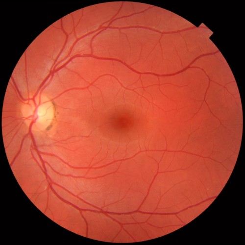
Fundus photography is the creation of a photograph of the interior surface of the eye, including the retina, optic disc, macula, and posterior pole. This instrument is used by eye professionals for monitoring progression of a disease, diagnosis of a disease (combined with retinal angiography), or in screening programs and epidemiology.
Compared to ophthalmoscopy, fundus photography generally needs a considerably larger instrument, but has the advantage of forwarding the image to a specialist at another location and/or time, as well as providing photo documentation for future reference. Modern fundus photographs generally recreate considerably larger areas of the fundus than what can be seen at any one time with handheld ophthalmoscopes.